eISSN:2278-5299

International Journal of Latest Research in Science and Technology

DOI:10.29111/ijlrst   ISRA Impact Factor:3.35

A News Letter Sign UP!
IN VITRO NEW BIOPOLYMER FOR BONE GRAFTING AND BONE CEMENT

Research Paper Open Access

International Journal of Latest Research in Science and Technology Vol.4 Issue 2, pp 46-55,Year 2015

IN VITRO NEW BIOPOLYMER FOR BONE GRAFTING AND BONE CEMENT

Gehan A. Raouf,Hana Gashlan, Alaa Khedr, Salem Hamedy, Hind Al-jabbri

Correspondence should be addressed to :

Received : 05 April 2015; Accepted : 08 April 2015 ; Published : 30 April 2015

Share
Download 125
View 179
Article No. 10490
Abstract

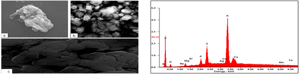
Cements based on synthetic calcium sulfate is among the most investigated material for dental and orthopedic applications in reconstructive surgery. To overcome the short-term cytotoxic effect, brittleness and fast resorption of calcium sulfate dihydrate(CSD), as bone substitute material, CSD was doped with Lepidium sativum water extract(LS) powder and mixed with cow bone mineral (BM) (1: 0.04: 1: wt/wt ratio respectively). The Fourier Transform Infrared (FTIR) spectra of BM-CSD, BM-CSD-LS, and BM-LS composites, compared to BM, were recorded. LS was analyzed by using Gas Chromatography/ Mass Spectroscopy(GC/MS) and Scanning Electron Microscope-Energy Disspersive X-ray(SEM-EDX). The results revealed that the total carbonate/phosphate ratio, type B-carbonate substitution, and acid phosphate content increased dramatically in all tested composites, the maximum increase was detected in BM+CSD+LS composite. Other forms, rather than B-carbonate substitution, takes place such as substitution with Zn, Mg and amino acids. The BM-crystallinity (BMC) decreased significantly in both BM+CSD, B+CSD+LS while, it is slightly decreased in BM+LS composite compared to BMC. Thus, addition of CSD to bone mineral leads to decrease in crystallinity while adding LS only or doping CSD with LS increases the crystallinity of bone mineral compared to BM+CSD composite with increasing in the apatite crystal size and the acid phosphate content as well.

Key Words   
Bone Mineral Crystallinity, Bone grafting, Calcium Sulphate Dihydrate, Fourier Transform Infrared
Copyright
References
  1. Athanasiou, K. A., C. F. Zhu, D. R. Lanctot, C. M. Agrawal, andX. 2000. Fundamentals of biomechanics in tissue

engineering of bone. Tissue Engineering. 6, 361-381.

  1. Lodish, H. 1995. Molecular Cell Biology Scientific American Books. New York.
  2. Demirkiran, H. 2012. Bioceramics for osteogenesis, molecular and cellular advances. Adv Exp Med Biol. 760, 134-47.
  3. Meunier, P. J., and J. F. Brantus. 1999. Losteoporose Le quotidien du medecin.
  4. Weiner, S., W. Traub.1992. Bone structure: from angstroms to microns. Faseb. 6, 879-885.
  5. de Groot, K. 1983. Ceramic of calcium phosphate: Preparation and properties; CRC Press Inc.
  6. Bajpai, P. K. 1992. A novel device for sustained long term delivery of drugs. Bioceramics 3, 87–89.
  7. Herve, P., V. Veronique, B. Wassila, M. Alain, de P. Cindy, B. Marianne, O. Karim, S. Laurent, and G. Genevieve. 2000. Tissue-engineered bone regeneration. Nature Biotechnology. 18, 959–963.
  8. Greenwald, A. S., S. D. Boden, V. M. Goldberg, Y. Khan, C. T. Laurencin, and R. N. Rosier, The Committee on Biological Implants.Bone-graft substitutes: facts, fictions, and applications. J.Bone Joint Surg Am. 83, 98-103.
  9. Kelly, C. M., R. M Wilkins, S. Gitelis, C. Hartjen, J. T. Watson, and P. T. Kim. 2001. The use of a surgical grade calcium sulfate as a bone graft substitute: results of a multicenter trial. Clin Orthop 382, 42-50.
  10. Pecora, G., D. De Leonardis, and J. Ricci. 2001. L’uso del solfato di Calcio Nelle Tecniche Rigenerative Multidisciplinari. Edizioni Elite Service, 13–77.
  11. Murashima, Y., G.Yoshikawa, R. Wadachi, N. Sawada, and H. Suda. 2002. Calcium sulphate as a bone substitute for various osseous defects in conjunction with apicectomy. Int. Endod 35, 768.
  12. Strocchi, R., G. Orsini, A. Iezzi, C. Scarano, G. Rubini, A. Pecora, and J. Piattelli. 2002. Bone regeneration with calcium sulfate: evidence for increased angiogenesis in rabbits. Oral Implantology 18, 273.
  13. Bell, W. H. 1964. Resorption rates of bone and bone substitutes. Oral Surg. 17, 650–7.
  14. Coetzee, A. S.1980. Regeneration of bone in the presence of calcium sulphate. Arch. Otolaryngol. 106, 405–9.
  15. Goren, S, H. Gokbayrak, and S. Altintas. 2004. Production of hydroxylapatite from animal bone. KEY ENGINEERING MATERIALS, 264-268: 1949-1952.
  16. Eddouks, M., M. Maghrani, N. A Zeggwagh, and J.B. Michel. 2005. Study of the hypoglycaemic activity of lepidium sativum l. Aqueous extract in normal and diabetic rats. J. Ethano. Pharmacol 97, 391–395.
  17. Bull, M., K. Manoj, H. K. Varma, and R. Sivakumar. 2000. On the development of an apatitic calcium phosphate bone cement. Biomedical Technology Wing. 23, 135–140.
  18. Forina, M., C. Armanino, and V. Raggio. 2002. Clustering with dendrograms on interpretation variables. Analytica Chimica Acta 454: 13-19.
  19. Abeysekara, S., D. Damiran, and P. Yu. 2013. Spectrochimica Acta - Part A: Molecular and Biomolecular Spectroscopy.102, 432-442.
  20. Nancy, P., B. Adele, and M. Richard. 1991. Novel infrared spectroscopic method for the determination of crystallinity of hydroxyapatite minerals. Biophysical Society. 786-793.
  21. Gunasekaran, K. and S.Thotapallip. 2011. Preparation and characterization of a novel bone graft composite containing bone ash and egg shell powder. Indian Academy of Sciences. 34, 177-181.
  22. Su-Gwam, K., K. Hak-Kyun, and L. Sung-Chul. 2001. Combined implantation of particulate dentine, plaster of Paris, and a bone xenograft (Bio-Oss1) for bone regeneration in rats. CranioMaxillofac Surg Clin North Am 29, 282–288.
  23. Navarro, M., S. del Valle, S. Martinez, S. Zeppetelli, L. Ambrosio, J. A Planell, and M. P. Ginebra. 2004. New macroporous calcium phosphate glass ceramic for guided bone regeneration. Biomaterialsn 25, 4233–4241.
  24. Thomson, R., M. J. P., Yaszemsk, J.M. owers, and A.G. Mikos. 1994. Fabrication of biodegradable polymer scaffolds to engineer trabecular bone. Biomater Sci Polym.7, 23–38.
  25. Giovanna, G., D. Alfredo, L. Paola, and M, Mario. 2007. Development of a new calcium sulphate-based composite using alginate and chemically modified chitosan for bone regeneration. Wiley Inter Science 34.
  26. Damien, C. J. and J. R. Parsons. 1991. Bone grafts and bone graft substitutes: A review of current technology and applications. Appl Biomater 2, 187–208.
  27. Ilan, D. I. and A. L. Ladd. 2003. Bone graft substitutes. Operat Tech Plast Reconstr Surg 9, 151–160.
  28. James, D. K., Y. Simon, K. Leda, W. Mark, and G. M. Antonios. 2009. Injectable Biomaterials for Regenerating Complex Craniofacial Tissues. Adv Mater 21, 3368–3393.
  29. Ricci, J. L., H. Alexander, P. Nadkarni, M. Hawkins, J.Turner, S. Rosenblum, L. Brezenoff, D. De Leonardis, and G. Pecora. 2001. Biological Mechanisms of Calcium Sulfate Replacement by Bone. Bone engineering. 332.
  30. Carinci, F., A. Piattelli, G. Stabellini, A. Palmieri, L. Scapoli, G. Laino, S. Caputi, and F. Pezzetti. 2004. Calcium sulfate: Analysis of MG63 osteoblast-like cell response by means of a microarray technology. Biomedical Materials Research 71B, 260.
  31. Walters, M. A., Y.C. Leung, N. C. Blumenthal, R. Z LeGeros, and K. AKonsker. 1990. A raman and infrared spectroscopic investigation of biological hydroxyapatite. Inorg Biochem 39, 193–200.
  32. Rey, C., B. Collins, T. Goehl, I. R. Dickson, and M. J. Glimcher. 1989. The carbonate environment in bone mineral: A resolutionenhanced Fourier transform infrared spectroscopy study. Calcif Tissue Int 45, 157–164.
  33. Bailey, R. T. and C. Holt. 1989. Fourier transform infrared spectroscopy and characterization of biological calcium phosphates. Macmillan Press. London.
  34. Legeros, R. Z. 1981. Apatites in Biological Systems. Prog. Crystal. Growth Charact. 4, 1-45.
  35. Yang, E. P., W. E. PASCHALIS, A.L. MAYO, A. Boskey, and R. Mendelsohn. 2001. Infrared Microscopic Imaging of Bone: Spatial Distribution of CO3. Bone and Mineral Research. 16.
  36. Elliott, J. C.1962. The infrared spectrum of the carbonate ion in carbonate-containing apatites. Dent Res 30, 1284.
  37. Emerson, W. H, and E. E. Fischer. 1962 The infra-red absorption spectra of carbonate in calcified tissues. Arch Oral Biol. 7, 671-683.
  38. Bonel, G. 1972. Contribution a l'etude de la carbonation des apatites. Part I. Ann. Chim 7, 65-87.
  39. Thompson, D. D., W. S. Posner, A. N. C. Laughlin, and N. C. Blumenthal. Comparison of bone apatite in osteoporotic and normal eskimos. Tissue Int 35, 392-393.
  40. Pleshko, N., A. Boskey, and R. Mendelsohn. 1991. Novel infrared spectroscopic method for the determination of crystallinity of hydroxyapatite minerals. Biophys 60, 786–793
  41. Paschalis, E. P., F. Betts, R. Mendelsohn, and A. L. Boskey. 1996. Fourier transform infrared spectroscopy of the solution-mediated conversion of amorphous calcium phosphate to hydroxyapatite: new correlations between X-ray diffraction and infrared data. Calcif. Tissue 58, 9–16.
  42. Adele, L. and M. Richard. 2005. Infrared spectroscopic characterization of mineralized tissues. Vib Spectrosc 38, 107–114.
  43. Elisa, B., T. Paola, G. Massimo, G. Roberto, and B. Adriana. 2006. Nanocomposites of hydroxyapatite with aspartic acid and glutamic acid and their interaction with osteoblast-like cells Chair of Surgical Pathophysiology 25, 4428-4433.
  44. Stewart, S. D. A., C. P. Shea, M. D. Tarnowski, D. Morris, and R. Wang. 2002. Trends in early mineralization of murine calvarial osteoblastic cultures: a Raman microscopic study. Raman Spectrosc 33, 536–543.
  45. Raisz, L. G. 1992. Mechanisms and regulation of bone resorption of osteoclastic cells. Raven Press, 287-311.
  46. Bart, C. 2008. Normal Bone Anatomy and Physiology. Clin J Am Soc Nephrol. S131–S139.
  47. Jitomir, J. and D. S. Willoughby. 2008. Leucine for retention of lean mass on a hypocaloric diet. Leucine for retention of lean mass on a hypocaloric diet. 11, 606-9.
  48. Yamaguchi, M. and R.Yamaguchi.1986. Action of zinc on bone metabolism in rats: Increase in alkaline phosphatase activity and DNA content. Biochem Pharmacol. 35, 773–777.
  49. Yamaguchi, M., H. Oishi, and Y. Suketa. 1987. Stimulatory effect of zinc on bone formation in tissue culture. Biochem Pharmacol. 36, 4007–4012.
  50. Yamaguchi, M., H. Oishi, and Y. Suketa.1988. Zinc stimulation of bone protein synthesis in tissue culture. Biochem Pharmacol. 37, 4075–4080.
  51. Saeed, H., N. Roghayeh, and N. Hamid, N. 2009. Physico–Chemical and In Vitro Biological Study of Zinc-Doped Calcium Sulfate. Wiley InterScience
  52. Moonga, B. S. and D. W. Dempster. 1995. Zinc is a potent inhibitor of osteoclastic bone resorption in vitro. Bone Miner Res. 10, 53–457
  53. Hashizume, M. and M. Yamaguchi. 1993. Stimulatory effect of beta-alanyl-L-histidinato zinc on cell proliferation is dependent on protein synthesis in osteoblastic MC3T3-E1 cells. Mol Cell Biochem. 122, 59–64.
  54. Hashizume, M. and M. Yamaguchi.1994. Effect of beta-alanyl-L-histidinato zinc on differentiation of osteoblastic MC3T3-E1 cells: increases in alkaline phosphatase activity and protein concentration. Mol Cell Biochem. 131, 19–24.
  55. Stein, G. S., J. B. Lian, and T. A. Owen. 1990. Relationship of cell growth to the regulation of tissue-specific gene expression during osteoblast differentiation. Faseb. 4, 3111–3123.
  56. Franceschi, R. T. and B. S. Iyer.1992. Relationship between collagen synthesis and expression of the osteoblast phenotype in MC3T3-E1 cells. Bone Miner Res. 7, 235–246.
  57. Coen, G., P. Ballanti, and E. Bonucci. 1998. Bone markers in the diagnosis of low turnover osteodystrophy in haemodialysis patients. Nephrol Dial Transplant. 13, 2294–302.
  58. Kamimura, H., N. K. O. Koga,H. Yoshimura,H. Inoue,K. Sato, and M. Ohkubo. 1989. Studies on distribution, excretion and subacute toxicity of squalene in dogs. Fukuoka Igaku Zasshi. 80, 269-280.
  59. Sandar , B. G. L., Z. M. Svetomir, I. Suzana, A. M. Dimitrijevi, andU. S. Dejan. 2007. Antimicrobial activity of the essential oil and differentfractions of juniperus communis l. and a comparison with some commercial antibiotics, Serbian Chemical Society. 72: 311-320.
  60. Lai, Y. L. and M.Yamaguchi. 2009. Phytocomponent p-hydroxycinnamic acid stimulates bone formation and inhibits bone resorption in rat femoral tissues in vitro. Mol Cell Biochem. 292, 45-52.
  61. Kotha, S. P., M. Lieberman, A. Vickers, S. R. Schmid, and J. J. Mason. 2006.  Adhesion enhancement of steel fibers to acrylic bone cement through a silane coupling agent. J Biomed Mater Res A. 76, 111-9.
  62. Nikolaos, K. and T. Margaret. 2010. Spectroscopic assessment of normal cortical bone: Differences in relation to bone site and sex, Scientific World. 10: 402–412.
  63. Nyquist, R. A., C. L. Putzig, and M. A. Leugers. 1997. The handbook of infrared and raman spectra of inorganic compounds and organic salts Edited by: San Diego, USA: Academic Press.
To cite this article

Gehan A. Raouf,Hana Gashlan, Alaa Khedr, Salem Hamedy, Hind Al-jabbri , " In Vitro New Biopolymer For Bone Grafting And Bone Cement ", International Journal of Latest Research in Science and Technology . Vol. 4, Issue 2, pp 46-55 , 2015


Responsive image

MNK Publication was founded in 2012 to upholder revolutionary ideas that would advance the research and practice of business and management. Today, we comply with to advance fresh thinking in latest scientific fields where we think we can make a real difference and growth now also including medical and social care, education,management and engineering.

Responsive image

We offers several opportunities for partnership and tie-up with individual, corporate and organizational level. We are working on the open access platform. Editors, authors, readers, librarians and conference organizer can work together. We are giving open opportunities to all. Our team is always willing to work and collaborate to promote open access publication.

Responsive image

Our Journals provide one of the strongest International open access platform for research communities. Our conference proceeding services provide conference organizers a privileged platform for publishing extended conference papers as journal publications. It is deliberated to disseminate scientific research and to establish long term International collaborations and partnerships with academic communities and conference organizers.